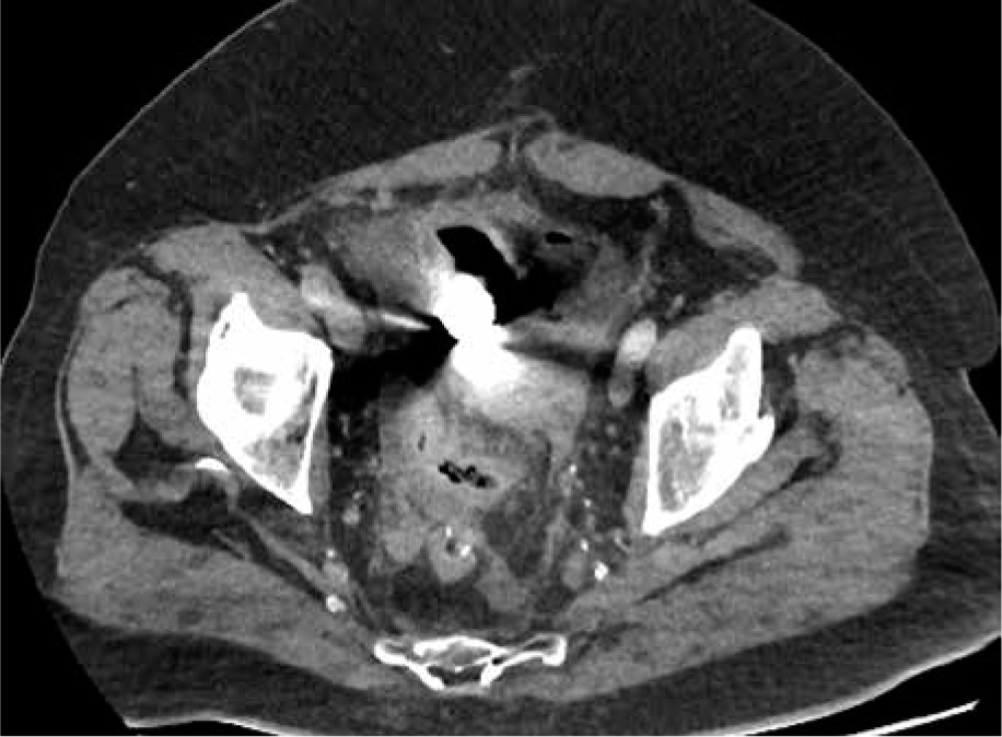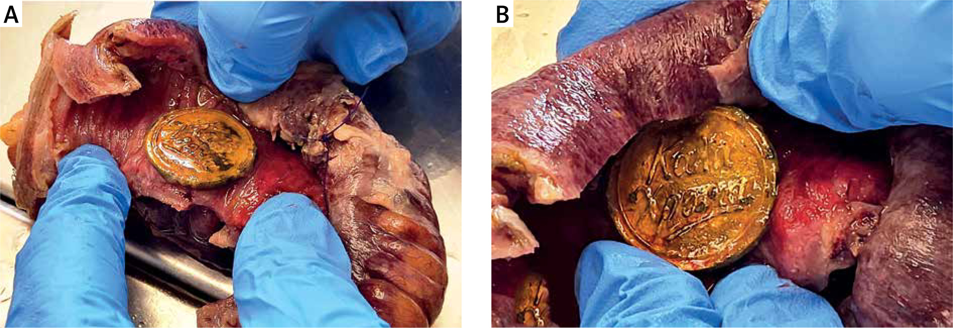


A 73-year-old male patient presented to the emergency department with diffuse severe abdominal pain persisting for 2 days. Upon clinical examination, the patient exhibited diffuse abdominal tenderness with signs of resistance and spasm, particularly in the left lower quadrant. Patient had undergone surgery for left colectomy due to colorectal cancer and subsequently received adjuvant chemotherapy, remaining disease-free to date. Additionally, the patient had a history of an inguinal hernia on the right side. The patient was haemodynamically unstable and oliguric at admission, necessitating vasoconstrictors. The patient’s vital signs revealed a blood pressure of 83/59 mm Hg and a pulse rate of 127 bpm. His blood pressure stabilized prior to computed tomography (CT) by receiving continuous IV fluid infusion. An abdominal CT scan revealed free intraperitoneal air and a foreign body within the gastrointestinal tract (Figure 1). Subsequently, the patient was taken to the operating room for exploratory laparotomy. Intraoperatively, faecal peritonitis and perforation of a segment of the terminal ileum were identified. A partial segmentectomy with simultaneous anastomosis was performed. During the resection of the surgical specimen, a medal trinket was discovered, originating from a New Year’s Eve cake (Figures 2 A, B). Following surgery, the patient was transferred to the intensive care unit, intubated, and mechanically ventilated. He remained hospitalized in the ICU for 8 days with no major postoperative complications. After a 21-day stay the patient was discharged. Despite the relatively common occurrence of ingesting foreign objects, most cases result in the object passing harmlessly through the alimentary tract without complications. Less than 1% of ingested foreign bodies lead to bowel perforation, which underscores the rarity of this outcome [1, 2]. While injuries resulting from ingested foreign bodies can manifest at various points along the digestive tract, the most common sites for perforation are found in the ileal, ileocaecal, and rectosigmoid regions [2–7]. Hence, a thorough understanding of the risks associated with ingested foreign bodies and their potential impact on the digestive system is crucial for both medical professionals and the general public, because it can aid in early recognition and prompt intervention when such cases arise [4, 5]. Foreign bodies that seem to be dangerous because of their shape and size should be effectively removed. There are many options available, including endoscopy, laparoscopy, and laparo-endoscopic removal of foreign bodies. The procedure of choice is endoscopic removal, which is fruitful in the majority of patients [8–10]. In conclusion, the perforation of intestinal structures due to ingested foreign bodies presents a complex and often challenging diagnostic scenario, especially in cases of acute abdominal symptoms, in which the underlying cause may not be immediately apparent. Abdominal CT scans can effectively identify instances of small bowel perforation and may provide valuable indications of the underlying cause [10]. Sometimes the laparoscopic approach before open surgery can be performed safely for the removal of a foreign body. Minimally invasive laparoscopic surgery should be preferred to open surgery due to its advantages [11, 12].
Czasopisma
Wszystkie czasopisma
Advances in Dermatology and Allergology/Postępy Dermatologii i Alergologii
Advances in Interventional Cardiology/Postępy w Kardiologii Interwencyjnej
Anaesthesiology Intensive Therapy
Archives of Medical Science
Biology of Sport
Central European Journal of Immunology
Folia Neuropathologica
Journal of Contemporary Brachytherapy
Pediatric Endocrinology Diabetes and Metabolism
Pielęgniarstwo Chirurgiczne i Angiologiczne/Surgical and Vascular Nursing
Polish Journal of Pathology
Prenatal Cardiology
Videosurgery and Other Miniinvasive Techniques